
A woman in her late thirties may notice the change while reorganizing her bra drawer after a second pregnancy. The cups still fit, but her favorite tops sit differently. The upper part of the breast looks less full, the nipple seems lower, and the skin near the bottom appears to gather above the crease beneath the breast. Nothing happened overnight. Her breast shape changed gradually through pregnancy, lactation, weight fluctuations, hormonal shifts, and aging.
That experience is common, but it doesn't automatically tell you whether you have ptosis of the breasts, how advanced it is, or which procedure would make sense. Ptosis is a medical description of breast descent, not a judgment about your body. The useful questions are where the nipple sits, how much excess skin is present, how much breast volume remains, and whether the breast has enough internal support to hold a new shape.
Table of Contents
- When Breasts Begin to Change Shape
- What Ptosis of Breasts Actually Means
- Why Breasts Sag and What Speeds It Up
- How a Specialist Measures and Grades Ptosis
- What Non-Surgical Options Can and Cannot Do
- Surgical Lift Options and How They Are Chosen
- Combining a Lift With Implants or Reduction
- Risks, Recovery, and How Long Results Last
When Breasts Begin to Change Shape
Breast shape develops through several overlapping stages. Puberty establishes the initial volume and skin envelope. Hormonal cycles can temporarily change fullness and tenderness. Pregnancy expands the glandular tissue and skin, and lactation adds another period of volume change before the breast may settle with less upper-pole fullness. Later, weight changes and menopause can alter the balance between glandular and fatty tissue.
The first signs are often visual rather than painful. You may notice:
- A lower nipple position: The nipple-areola complex sits closer to, or below, the fold beneath the breast.
- A flatter upper pole: The area above the nipple loses the fullness that once made the breast look rounder.
- Lower-pole skin laxity: The skin stretches downward and may appear to pool just above the inframammary fold.
- A different clothing fit: The breast may occupy more space in the lower cup without adding the fullness you want near the collarbone.
These changes reflect the way breast tissue is supported. The skin envelope gradually loses elasticity, glandular volume can deflate, and the internal suspensory structures carry the effects of years of breast weight. Pregnancy and substantial weight change can make the contrast more noticeable because the skin expands and then has less tissue volume to contain.
A useful distinction: Breast volume and breast position aren't the same problem. An implant can add volume, but it may not correct a nipple that has descended below the breast fold.
Breast ptosis has been formally classified in modern plastic surgery for decades. The Regnault grading system was introduced in 1976 and remains a standard reference for describing severity, as outlined in this history of breast lift classification. Understanding that vocabulary makes a consultation more productive because you can discuss shape in measurable terms rather than relying only on words such as “sagging” or “deflated.”
What Ptosis of Breasts Actually Means
Think of a balloon held inside a mesh net. If the net becomes slack, the balloon settles lower even if the balloon itself hasn't changed dramatically. Breast ptosis follows a similar mechanical pattern. The skin envelope and internal support become less effective, and the breast tissue descends.
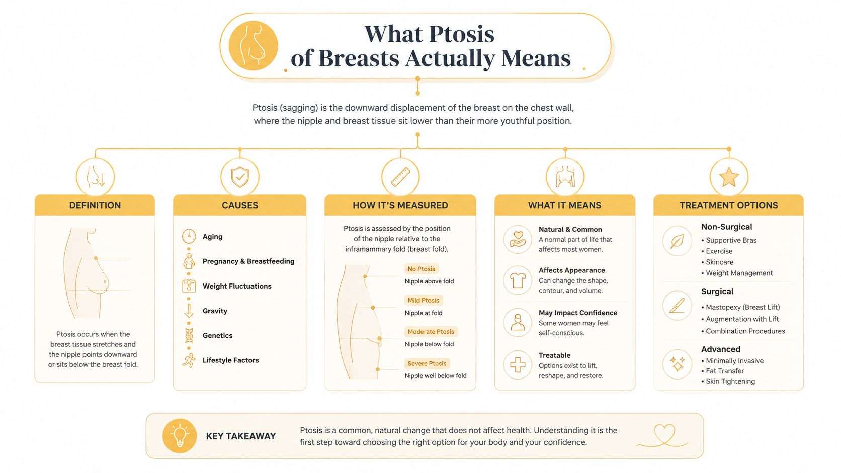
Clinically, the key reference point is the inframammary fold, the natural crease where the lower breast meets the chest wall. A specialist compares the position of the nipple-areola complex with that fold while the breast is assessed at rest.
The Regnault grades in plain language
- Grade I, or mild ptosis: The nipple sits at the level of the inframammary fold. A related published description places the nipple at, or up to 1 cm below, the fold, depending on the classification used. The breast may have mild lower-pole looseness while retaining a relatively stable overall contour.
- Grade II, or moderate ptosis: The nipple sits below the fold but remains above the lowest point of the breast mound. In a separate published framework, this corresponds to the nipple sitting 1 to 3 cm below the fold. The nipple usually still points forward.
- Grade III, or severe ptosis: The nipple reaches the most dependent portion of the breast and may point downward. The same framework describes this as more than 3 cm below the fold or at the inferior pole. The skin envelope typically has more substantial redundancy.
A prospective anthropometric study using the Regnault grades similarly defined Grade I as the nipple descending to the fold, Grade II as below the fold but above the lowest contour, and Grade III as below the fold at the lowest contour, as shown in this prospective breast measurement study.
Pseudoptosis can confuse the picture. In pseudoptosis, the nipple may remain near the fold while the lower breast tissue hangs below it. Breast size, implant history, skin quality, and the distribution of glandular and fatty tissue all affect the appearance, so a photograph or bra fit can't establish the grade by itself.
Why Breasts Sag and What Speeds It Up
The breast isn't a muscle that can be tightened through chest exercises. Its shape depends on a skin envelope, glandular and fatty tissue, fibrous support such as Cooper's ligaments, and the way the breast attaches across the chest wall. The pectoral fascia provides an important deep reference layer, but it doesn't function like a taut internal bra that can prevent every change.
The structures under stress
Pregnancy expands the glandular component and stretches the skin. After lactation and weaning, the glandular volume may reduce, leaving the envelope relatively loose. Weight cycling adds another type of strain because the skin repeatedly expands and contracts, while a large reduction in volume can leave more skin than the breast can fill.
Aging gradually changes collagen and elastin quality. Gravity then acts on the breast's weight every day, and the effect becomes more visible when the skin and internal support have already lost elasticity. Hormonal changes around menopause can also shift the proportion of glandular tissue and fat, often making the upper breast appear less full.
Research has identified several meaningful risk factors. A case-control study found that age, BMI, a history of weight loss greater than 50 pounds, larger bra cup size, number of pregnancies, and smoking history were significant risk factors, while breastfeeding itself wasn't a significant predictor, according to this clinical overview of breast ptosis. That distinction matters. A patient shouldn't assume that lactation alone caused the change, or that avoiding breastfeeding would have prevented it.
| Cause | Primary structure affected | Typical result on shape |
|---|---|---|
| Pregnancy and postpartum volume change | Skin envelope and glandular tissue | Expanded skin followed by reduced fullness and lower-pole descent |
| Significant weight loss | Skin envelope and soft-tissue volume | Redundant skin with less tissue filling the upper pole |
| Aging | Collagen, elastin, and suspensory support | Gradual descent and reduced firmness |
| Larger breast volume | Skin and internal supporting structures | Greater gravitational load and lower-pole stretch |
| Smoking history | Skin quality and healing capacity | Weaker elasticity and greater concern during surgical healing |
| Hormonal change | Glandular-to-fat balance | A softer, less full upper breast |
You can influence some contributors, particularly weight stability and smoking cessation. Sun protection may support general skin health, but creams and lifestyle changes can't remove stretched skin or reposition a descended nipple. Those structural changes require an individualized discussion of Breast Lift, Super Breast Lift, Breast Reduction, or another approved breast procedure when appropriate.
How a Specialist Measures and Grades Ptosis
A breast consultation begins with observation, but it shouldn't end there. A specialist examines the breast without relying on how it looks inside a bra. The patient stands naturally while the surgeon evaluates nipple position, the breast fold, asymmetry, skin quality, breast base, and how much tissue sits above and below the nipple.
The measurements that shape the plan
The suprasternal notch-to-nipple distance measures the vertical relationship between the notch at the base of the neck and the nipple. It helps compare breast height and asymmetry. The nipple-to-inframammary-fold relationship shows how far the nipple has descended relative to the lower breast crease and helps characterize the lower-pole skin excess.
A surgeon may also record:
- Breast base width: This helps determine how much breast footprint exists on the chest wall and whether an implant could fit without creating excessive width.
- Volume distribution: A breast with adequate lower-pole tissue but little upper-pole fullness may require a different strategy from a breast with substantial excess volume.
- Skin elasticity: Stretch and recoil help predict whether the skin can support an implant or whether a lift must carry more of the correction.
- Asymmetry: Differences in nipple height, fold position, volume, or chest-wall shape may require separate adjustments on each side.
The Regnault grade provides a shared language, but it doesn't dictate one operation. A Grade I breast with poor elasticity may need more support than a Grade II breast with better tissue quality. Published planning frameworks have also used anterior pull skin stretch under 4 cm and nipple-to-inframammary-fold distance under maximal stretch under 10 cm as thresholds compatible with moderate laxity for selected augmentation strategies. Values above those limits suggest reduced elasticity and a stronger indication for mastopexy-based correction, as described in this breast augmentation and mastopexy planning chapter.
The measurements don't predict an identical result for every patient. They help the surgeon decide how much skin needs adjustment, whether the nipple needs repositioning, and whether Breast Augmentation would add useful volume or place more weight on a weak envelope.
What Non-Surgical Options Can and Cannot Do
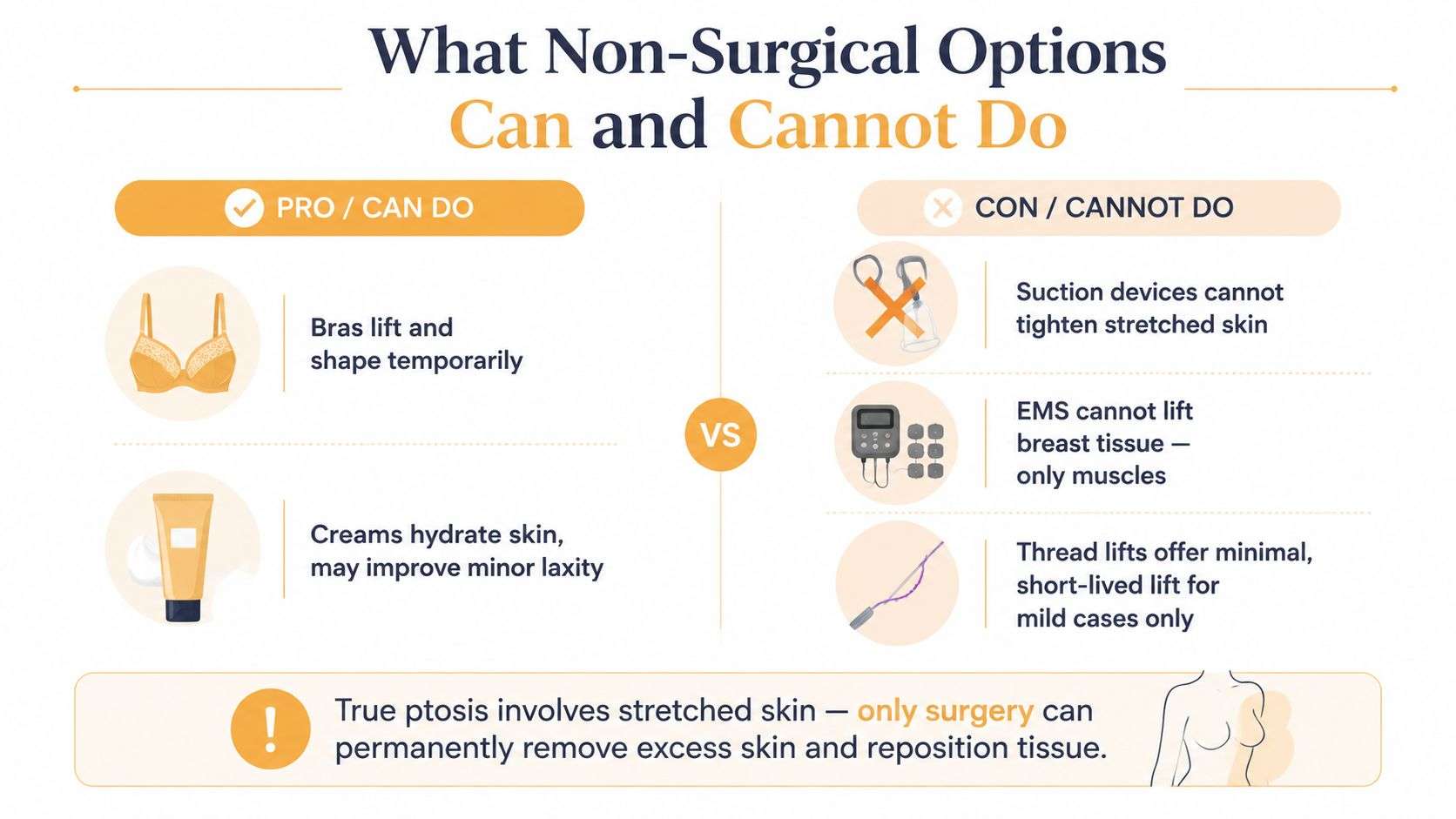
Many patients search for a way to correct breast ptosis without surgery. That preference is understandable. The difficulty is that true ptosis involves stretched skin, weakened internal support, and descended breast tissue. A topical product can't remove excess skin, and an exercise program can't move the nipple-areola complex to a higher position.
A supportive bra can still be useful. It can improve comfort, reduce movement during activity, and create a higher silhouette under clothing. Weight stabilization may also reduce additional stretching. Those measures manage appearance and comfort, but they don't reverse established descent.
Separating temporary support from structural correction
Creams can hydrate the surface of the skin and may improve its feel. They can't rebuild the breast's suspensory anatomy. Suction devices may temporarily alter fluid distribution or create a short-lived visual effect, but they don't reliably tighten a stretched envelope. Electrical muscle stimulation affects muscle, not the breast tissue itself.
Minimally invasive energy-based approaches have been described as producing only moderate improvement in mild to moderate ptosis, and the available classification and assessment literature remains limited, with one systematic review identifying 16 observational studies and 2 randomized studies, according to this review of breast ptosis. That evidence gap is important for anyone considering a lower-downtime treatment. Modest skin firming isn't equivalent to nipple repositioning or removal of redundant skin.
A thread lift may offer limited, short-lived elevation in selected mild cases, but it doesn't provide the same structural correction as a mastopexy. Patients with more pronounced Grade II or Grade III ptosis should approach nonsurgical marketing cautiously and discuss whether an operation is the more realistic path.
For a patient focused on lower-downtime choices, the discussion of natural breast lift options can help clarify what nonoperative measures may improve and where their limits begin. The decision should still be based on an examination, not an advertisement or a self-assigned grade.
Surgical Lift Options and How They Are Chosen
A mastopexy changes the relationship between the nipple, breast mound, and skin envelope. The incision pattern is selected according to the amount of descent, the quantity of excess skin, the size and position of the areola, the elasticity of the tissue, and whether the patient wants added volume or reduced volume.
Matching the pattern to the problem
A crescent lift removes a small amount of skin above the areola and suits carefully selected mild cases with limited nipple repositioning. A periareolar, or “donut,” lift places the scar around the areola and can address mild descent or areolar enlargement, although excessive tension can flatten the breast or widen the scar in patients with poor skin quality.
The vertical, or lollipop, pattern adds a line from the areola toward the fold. It allows the surgeon to reshape the lower pole and manage more moderate descent without extending the scar fully across the crease. An inverted-T, or anchor, pattern adds a horizontal incision in the fold. It offers the greatest access for significant skin removal and is often considered for advanced Grade III ptosis or substantial lower-pole redundancy.
The Super Breast Lift takes a different approach to support. Rather than depending on skin tension alone, it uses internal suspension to redistribute glandular weight toward the chest wall. That concept is intended to place less strain on the skin envelope and can be considered when long-term shape retention is a central concern.
| Technique | Best ptosis grade | Scar pattern | Ideal candidate |
|---|---|---|---|
| Crescent lift | Selected Grade I | Short crescent above the areola | Mild descent with limited repositioning |
| Periareolar lift | Grade I and selected mild Grade II | Around the areola | Mild laxity or areolar enlargement |
| Vertical lift | Grade II and selected Grade III | Around the areola and vertically downward | Moderate descent with lower-pole reshaping needs |
| Inverted-T lift | Advanced Grade III | Around the areola, vertical, and along the fold | Significant excess skin and lower-pole redundancy |
| Super Breast Lift | Selected mild to advanced cases | Determined by skin removal needs | Patients needing internal support as well as reshaping |
The trade-off patients should understand
Shorter scars aren't automatically better if they can't remove enough skin or support the desired shape. Longer scars can provide more control, while internal support may improve the way the breast carries its weight. Future pregnancy, anticipated weight change, breast size, and the patient's tolerance for scars all belong in the decision.
A lift can stand alone when the patient wants a higher, reshaped breast without more volume. If the upper pole has lost volume, the surgeon may discuss Breast Augmentation with the lift. If the breast is heavy or causing physical symptoms, Breast Reduction may be more appropriate. Patients considering a lift without added volume can review the approach to a breast lift without implants before discussing their anatomy in person.
Combining a Lift With Implants or Reduction
A lift with augmentation isn't two unrelated procedures performed at once. The implant adds weight and volume while the lift tightens and reshapes the skin envelope. The implant must fit the breast footprint and the newly raised tissue. Choosing too much volume for a thin or lax envelope can place more tension on the repair and may contribute to future descent or revision concerns.
The surgeon also considers the implant pocket, nipple position, breast-fold position, asymmetry, and skin stretch. In some patients, a combined operation is reasonable. In others, a staged plan is safer or more predictable, especially when the breast has severe laxity, very thin tissue, a previous lift that has stretched, implant malposition, bottoming out, capsular contracture, rupture, or visible rippling.
When reduction fits better
Reduction removes volume while lifting and reshaping the remaining tissue. The surgeon must preserve blood supply to the nipple-areola complex while choosing how much tissue to remove. A larger, heavier breast may need more skin management and a more supportive lift pattern, but the final decision depends on the breast footprint, tissue quality, symptoms, and goals.
Breast Implant Revision may include implant exchange, implant removal, pocket correction, or an exchange with lift when existing implants and ptosis are connected. Breast Implant Removal may be performed with or without a lift, depending on how much skin remains after the implant is removed and how high the patient wants the nipple positioned.
| Factor | Lift plus augmentation | Lift plus reduction |
|---|---|---|
| Main objective | Elevate tissue and restore or add volume | Elevate tissue while decreasing volume |
| Important planning issue | Implant size, pocket, skin elasticity, and footprint | Tissue removal, nipple blood supply, and final breast weight |
| Typical patient concern | Deflation or loss of upper-pole fullness | Excess weight, size, or lower-pole heaviness |
| Staging considerations | Thin tissue, severe laxity, or implant complications may alter timing | Major asymmetry or substantial tissue removal may affect the plan |
| Long-term trade-off | Added implant weight remains part of future shape changes | Reduced weight may lessen heaviness, but aging and weight change continue |
Patients often want to know whether results justify the recovery. A 2026 meta-analysis reported pooled satisfaction of 84.7%, while also linking preoperative anxiety and unrealistic expectations with lower satisfaction and more revision requests, as reported in this meta-analysis of mastopexy outcomes. The number is encouraging, but it doesn't promise an individual result. Candidacy, healing, and agreement about the desired size and shape matter just as much as the operation selected.
For patients considering volume restoration with elevation, the discussion of breast lift with augmentation can provide useful background before a consultation. An examination remains necessary to decide whether augmentation, reduction, revision, removal, or a lift alone best fits the anatomy.
Risks, Recovery, and How Long Results Last
A mastopexy creates scars, and scar placement depends on the amount of correction required. Patients should also discuss changes in nipple sensation, asymmetry, delayed healing, infection, bleeding, wound separation, changes in breastfeeding potential, and the possibility of revision. These risks vary with the technique, smoking history, tissue quality, medical history, implant status, and whether the lift is combined with Breast Augmentation, Breast Reduction, Breast Reconstruction, or implant revision.
Recovery isn't a single milestone. Early care may include a surgical bra, activity restrictions, incision monitoring, and medication instructions. Desk work often resumes before strenuous exercise, while lifting and upper-body exertion remain restricted until the surgical team confirms adequate healing. Swelling and firmness can make the breasts look temporarily uneven, and the final contour settles gradually.
A lift resets the breast position, but it doesn't stop aging, gravity, future pregnancy, or weight change. Long-term shape maintenance depends partly on the original skin quality and breast weight. The operation can improve descent, but it isn't a permanent freeze of the body.
Ptosis also matters in reconstruction. Contemporary oncoplastic planning uses the degree of ptosis as a technique-selection variable, and staged approaches can expand options for patients with moderate or severe descent. In staged-immediate reconstruction, sometimes called the Zenn delay, reconstruction typically begins about 2 to 3 weeks after mastectomy, and ptosis can be one reason for choosing that timing because it may help address skin-perfusion risk, as described in this review of staged-immediate breast reconstruction. Reconstruction after mastectomy therefore requires a separate conversation about cancer treatment, skin quality, nipple-sparing candidacy, and staging.
Schedule a consultation with OC Breast Surgery in Newport Beach to discuss breast ptosis, implant revision or removal, Breast Reduction, Super Breast Lift, Breast Reconstruction, and Breast Augmentation options with Dr. Mark Anton, the medical director and founder of OC Breast Surgery. Bring your goals, prior operative records, and questions about scars, recovery, future pregnancy, and whether a staged plan may be appropriate for your anatomy.